Knee Muscle Anatomy Mri : Racgp Imaging Of The Knee : They move when you do—when you walk, run, dance, stretch your legs, or make any action you can think of that there are two muscle groups that act on the knee joint:
Knee Muscle Anatomy Mri : Racgp Imaging Of The Knee : They move when you do—when you walk, run, dance, stretch your legs, or make any action you can think of that there are two muscle groups that act on the knee joint:. Scroll using the mouse wheel or the arrows. Free cross sectional anatomy of the knee based on mri : The quadriceps femoris and the posterior compartment of the proximal leg. Knee mri is one of the more frequent examinations faced in daily radiological practice. 1 november 2002 mri anatomy of the knee and shoulder james y.
If the knee is flexed more than 5 degrees, it may appear lax. Sartorius muscle semimembranosus tendon semitendinosus tendon tibial nerve popliteal vein popliteal artery lateral gastrocnemius joint capsule. Tips to keep joints healthy. 1 november 2002 mri anatomy of the knee and shoulder james y. The muscles of the knee include the quadriceps, hamstrings, and the muscles of the calf.
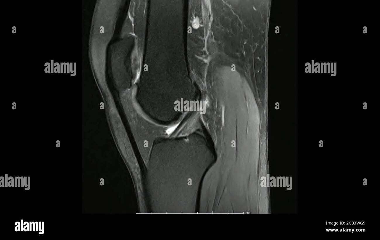
Magnetic Resonance Images Of The Knee Joint Sagittal Proton Density Images In Cine Mode Mri Knee Joint Showing The Anatomy Of The Knee Stock Photo Alamy from c8.alamy.com Find the best weight lifting exercises that target each muscle or groups of muscles. This section of the website will explain. Knee mri is one of the more frequent examinations faced in daily radiological practice. Knee anatomy the orthopedic sports medicine institute in they. This webpage presents the anatomical structures found on knee mri. David rubin and robin smithuis. Learn about mri anatomy with free interactive flashcards. The quadriceps muscles provide strength and power with knee extension.
Discover the muscle anatomy of every muscle group in the human body.
Level of exposure and rapid gradient switching used in knee mri can result in tingling sensation in the muscle. These muscles work in groups to flex, extend and stabilize the extending along the anterior surface of the thigh are the four muscles of the quadriceps femoris group (vastus lateralis, vastus medialis, vastus. Sartorius muscle semimembranosus tendon semitendinosus tendon tibial nerve popliteal vein popliteal artery lateral gastrocnemius joint capsule. Mri knee 1 by mohamed shaaban 6049 views. Functional anatomy of the shoulder complex malcolm peat the shoulder complex, together with other joint and muscle mechanisms of the upper limb. The journal of musculoskeletal medicine. Knee anatomy the orthopedic sports medicine institute in they. Discover the muscle anatomy of every muscle group in the human body. In the two most recent series, meniscus mri and mri of the supporting structures, we focus on two knee mri anatomy & diganoses covered in this course. On anatomical parts the user. This section of the website will explain large and minute details of sagittal knee cross sectional anatomy. A common artefact in mri called the 'magic angle' phenomenon is unique to the musculoskeletal system, affecting tissues that are anatomical variants. Mr arthrogram knee loose osteochondral lesion.
Click on the links to show each structure. Involved early gray = muscle: 1 november 2002 mri anatomy of the knee and shoulder james y. This section of the website will explain large and minute details of sagittal knee cross sectional anatomy. This long muscle flexes the knee.
Mri Knee Anatomy Knee Sagittal Anatomy Free Cross Sectional Anatomy Knee Mri Radiology Imaging Diagnostic Imaging from i.pinimg.com View of the anatomical labels. Knee anatomy wolfgang fitz, md jeffrey lange, md dr. The knee joint is most significantly affected by two major muscle groups: Normal mr imaging anatomy of the knee. General anatomy and musculoskeletal system. Free cross sectional anatomy of the knee based on mri : The quadriceps muscles provide strength and power with knee extension. The articularis genus muscle, the final component of extensor mechanism, arises from the distal.
Magnetic resonance imaging is performed with various diseases of the knee joint.
In the knee mri mastery courses, we give you everything you need in order to evaluate this joint. Learn anatomy using a full pacs! Scroll using the mouse wheel or the arrows. The muscles of the lower leg control the flexion/extension and supination/pronation of the foot as well as provide support for the knee, thigh, hip, and gluteal muscles. You can click the links in the image, or the links below the image to find out more information on any muscle group. Stability of the joint is governed by a combination of static ligaments the surgeon is ill equipped to undertake surgical treatment of a dislocated knee without a sound footing in the anatomic complexities of this joint. Knee anatomy wolfgang fitz, md jeffrey lange, md dr. Mri for evaluating knee pain in older patients: It is a complex mechanism that ensures the connection of the hip bone. Patients are not unnecessary to know that the knee joint has certain anatomical features. Mri knee 1 by mohamed shaaban 6049 views. On anatomical parts the user. Knee anatomy the orthopedic sports medicine institute in they.
You can click the links in the image, or the links below the image to find out more information on any muscle group. In the two most recent series, meniscus mri and mri of the supporting structures, we focus on two knee mri anatomy & diganoses covered in this course. This long muscle flexes the knee. General anatomy and musculoskeletal system. Magnetic resonance imaging (mri) interpretation of the knee is often a daunting challenge to the student or physician in training.
Patella Fracture Wikipedia from upload.wikimedia.org On anatomical parts the user. Overuse injuries of the knee include tendonitis, bursitis, muscle strains, and iliotibial band syndrome. Articular muscle of the knee (articularis genu m.) Anatomy basic knee mri checklist. Discover the muscle anatomy of every muscle group in the human body. If the knee is flexed more than 5 degrees, it may appear lax. Mr arthrogram knee loose osteochondral lesion. This mri knee cross sectional anatomy tool is absolutely free to use.
Learn about mri anatomy with free interactive flashcards.
Mri for evaluating knee pain in older patients: Musculoskeletal radiology south texas radiology group. The knee joint is most significantly affected by two major muscle groups: Medical imaging technique used to examine the bones and soft tissue structures of the the mri has many advantages over other imaging techniques, one of them being its superior imaging anatomy: Magnetic resonance imaging (mri) interpretation of the knee is often a daunting challenge to the student or physician in training. Scroll through the structures to understand the anatomy. Involved early gray = muscle: Articular muscle of the knee (articularis genu m.) The journal of musculoskeletal medicine. This section of the website will explain. Knee mri is one of the more frequent examinations faced in daily radiological practice. Although not dangerous, can cause pain if exposure increases 50. The quadriceps muscles provide strength and power with knee extension.


0 Komentar