Anatomy Of Chest Bones : The Bones Of The Chest And Upper Back Anatomy Medicine Com / A collection of anatomy notes covering the key anatomy concepts that medical students need to learn.
Anatomy Of Chest Bones : The Bones Of The Chest And Upper Back Anatomy Medicine Com / A collection of anatomy notes covering the key anatomy concepts that medical students need to learn.. Learn about chest anatomy with free interactive flashcards. Parts of the chest bones for many, the chest is made up of a. They are always longer than they are wide the vertebrae are irregular bones. Long bones are mostly located in the appendicular skeleton and include bones in the lower limbs (the tibia, fibula, femur, metatarsals, and phalanges) and bones in the upper limbs (the humerus, radius, ulna, metacarpals. It can help you understand our world more detailed and specific.
Bones have many shapes and sizes and are important to add structure to the body and protection to the vital structures. Bone of chest and their parts. Radiological anatomy of the chest please view our editing file before studying this lecture to check for any changes. Language and terminology for the study of the anatomy of the thorax. Learn about chest anatomy with free interactive flashcards.
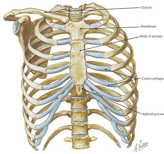
Thorax Wikipedia from upload.wikimedia.org The manubrium, sternal body, and xiphoid process. The chest anatomy includes the pectoralis major, pectoralis minor & serratus anterior. A collection of anatomy notes covering the key anatomy concepts that medical students need to learn. It, essentially, floats off of the back of the chest, as it is connected to the. The medial anterior chest is defined by the sternum, which consists of 3 flat polygonal bones: These joints fuse together in adulthood, thus permitting brain growth during adolescence. Long bones are mostly located in the appendicular skeleton and include bones in the lower limbs (the tibia, fibula, femur, metatarsals, and phalanges) and bones in the upper limbs (the humerus, radius, ulna, metacarpals. Long bones are categorised by their tubular shaft (diaphysis) with a rounded end (epiphysis) on each end.
Learn about each muscle, their locations & functional anatomy.
Learn about chest anatomy with free interactive flashcards. Ground substance and collagen fibers create a matrix that contains. All of the anatomical and important histological facts about the bones, together with the clinical relations, are going to be desrcibed in this article. Bones of the chest and upper back (posterior view). Reference database of normal imaging from birth to age 16. Bone basics and bone anatomy. It originates at your clavicle, ribs, and sternum, and inserts into the upper portion of your humerus (upper arm bone from elbow to shoulder.) Language and terminology for the study of the anatomy of the thorax. Bones have many shapes and sizes and are important to add structure to the body and protection to the vital structures. A collection of anatomy notes covering the key anatomy concepts that medical students need to learn. It extends from the position of the diaphragm to the clavicle or. The manubrium, sternal body, and xiphoid process. We hope you will use this picture in the study and helping chest and abdominal cavities with some organs removed.
Learn about each muscle, their locations & functional anatomy. Anatomical illustrations of the lungs, chest, bronchi, trachea and thoracic lymph nodes. Anatomy of the chest wall. It extends from the position of the diaphragm to the clavicle or. Reference database of normal imaging from birth to age 16.
Bones Of The Human Chest Front View Human Anatomy Medical Science Poster Stock Illustration Illustration Of Drawing Front 157797491 from thumbs.dreamstime.com They are always longer than they are wide the vertebrae are irregular bones. Clavicle bone is a narrow rod like s shaped bone extending between sternum and acromion process of scapula bone. Long bones are categorised by their tubular shaft (diaphysis) with a rounded end (epiphysis) on each end. Parts of the chest bones for many, the chest is made up of a. The bones have a the scapula, or shoulder blade, is an approximately triangular shaped bone. Atlas of anatomy of the human body: Science anatomy scan of human body organs and bones. When a patient flexes the neck forward, the prominent process is usually that of the 7th cervical.
All of the anatomical and important histological facts about the bones, together with the clinical relations, are going to be desrcibed in this article.
Computerized tomography 4 anatomy of lung segmental anatomy of lung lateral view on a normal lateral view the contours of the heart are visible and the ivc is seen entering the right atrium. Anatomy is the amazing science. It is comprised of many bones, formed by intramembranous ossification, which are joined together by sutures (fibrous joints). Breast bone anatomy human breast bone anatomy bone anatomy sternum | chest bone : Learn skull anatomy with skull bones quizzes and diagram labeling exercises. A collection of anatomy notes covering the key anatomy concepts that medical students need to learn. Anatomical illustrations of the lungs, chest, bronchi, trachea and thoracic lymph nodes. identify the bones of the thoracic cage. Learn about chest anatomy with free interactive flashcards. Human chest bone structure parts of the chest bones. Bone of chest and their parts. Chest bone, ribs, lung, heart, xiphoid process. All of the anatomical and important histological facts about the bones, together with the clinical relations, are going to be desrcibed in this article.
Bones of the chest and upper back (posterior view). Have you ever seen fossil remains of dinosaur and ancient human bones in textbooks, television, or in person at a museum? All of the anatomical and important histological facts about the bones, together with the clinical relations, are going to be desrcibed in this article. Ground substance and collagen fibers create a matrix that contains. When a patient flexes the neck forward, the prominent process is usually that of the 7th cervical.
Thorax Radiology Key from radiologykey.com Long bones are categorised by their tubular shaft (diaphysis) with a rounded end (epiphysis) on each end. Parts of the chest bones for many, the chest is made up of a. Science anatomy scan of human body organs and bones. A collection of anatomy notes covering the key anatomy concepts that medical students need to learn. Atlas of anatomy of the human body: All of the anatomical and important histological facts about the bones, together with the clinical relations, are going to be desrcibed in this article. Long bones function to support the weight of the body and facilitate movement. Bone of chest and their parts.
Anatomy is the amazing science.
We hope you will use this picture in the study and helping chest and abdominal cavities with some organs removed. Long bones function to support the weight of the body and facilitate movement. Ground substance and collagen fibers create a matrix that contains. It is comprised of many bones, formed by intramembranous ossification, which are joined together by sutures (fibrous joints). Anatomy is the amazing science. Anatomy of the chest wall. Your rib cage, for example, acts like a shield around your chest to protect important organs inside such as your lungs and heart. An overview of the anatomy of the hand, including the bones of the hand, muscles, blood supply and nerve supply. These joints fuse together in adulthood, thus permitting brain growth during adolescence. Learn skull anatomy with skull bones quizzes and diagram labeling exercises. The rib cage is one of the bodys best defenses against injury from impact. This webpage presents the anatomical structures found on wrist mri. Computerized tomography 4 anatomy of lung segmental anatomy of lung lateral view on a normal lateral view the contours of the heart are visible and the ivc is seen entering the right atrium.
The manubrium, sternal body, and xiphoid process anatomy of chest. Anatomy of the chest wall.


0 Komentar